Conjoint Tendon Shoulder Anatomy / 215. Interfoveolar ligament and conjoint tendon / Coracohumeral ligament middle glenohumeral ligament superior glenohumeral ligament long head of the biceps tendon anterior joint capsule.
Conjoint Tendon Shoulder Anatomy / 215. Interfoveolar ligament and conjoint tendon / Coracohumeral ligament middle glenohumeral ligament superior glenohumeral ligament long head of the biceps tendon anterior joint capsule.. Webmd's shoulder anatomy page provides an image of the parts of the shoulder and describes its the shoulder is one of the largest and most complex joints in the body. The conjoint tendon is a sheath of connective tissue that attaches the transversus abdominis, the deepest of the four abdominal muscles, to the pelvis. The shoulder anatomy includes the anterior, lateral & posterior deltoids, plus the rotator cuff. Shoulder joint allows lifting, pushing and pulling by upper extremity. Cal, cp and the conjoint tendon should be evaluated as an important osteotendinoligamentous arch supporting the shoulder joint anterosuperiorly.
The conjoint tendon, also known as the inguinal aponeurotic falx or henle's ligament, is a condensation of tissue that runs through the lateral edge of the conjoint tendon forms the medial part of the posterior wall of the inguinal canal.3 it is located right behind the superficial inguinal ring. The inguinal aponeurotic falx (falx aponeurotica inguinalis; Anterior graphic of the shoulder. They can withstand a degree of stretching and turning as tendon sheaths are located around tendons, which are found in joints throughout the body, including the hands, arms, shoulders, legs, and feet. Conjoined tendon of internal oblique and transversalis muscle) of the obliquus internus and transversus is mainly formed by the lower part of.
Abdominal Obliques: A Diagram of the Torso - Holistic ... from herniaremediation.org Tendon conjoint — le tendon conjoint ici noté inguinal aponeurotic falx le tendon conjoint est une structure fibreuse constitué de la réunion des terminaisons fibreuses des muscles oblique interne et transverse de l abdomen. The conjoint tendon is a sheath of connective tissue that attaches the transversus abdominis, the deepest of the shoulder anatomy is an elegant piece of machinery having the greatest range of motion of any joint in the body. The conjoint tendon (previously known as the inguinal aponeurotic falx) is a sheath of connective tissue formed from the lower part of the common aponeurosis of the abdominal internal oblique muscle and the transversus abdominis muscle, joining the muscle to the pelvis. Coracohumeral ligament middle glenohumeral ligament superior glenohumeral ligament long head of the biceps tendon anterior joint capsule. The purpose of this study was to determine the effectiveness of open conjoint tendon release in patients with anterior shoulder pain due to conjoint. The conjoint tendon can be describe as a layer of connective tissue which connects the pelvis to the transversus abdominis, the. Cal, cp and the conjoint tendon should be evaluated as an important osteotendinoligamentous arch supporting the shoulder joint anterosuperiorly. Shoulder joint allows lifting, pushing and pulling by upper extremity.
The tendon of the subscapularis muscle attaches both to the lesser tubercle aswell as to the greater tubercle giving support to the long head of the biceps in.
Cal, cp and the conjoint tendon should be evaluated as an important osteotendinoligamentous arch supporting the shoulder joint anterosuperiorly. Il rentre jeu dans la formation du… … wikipédia en français. The purpose of this study was to determine the effectiveness of open conjoint tendon release in patients with anterior shoulder pain due to conjoint. Learn their origins/insertions, functions & exercises. The ssc tendon contributes to the formation of an anatomical space called the rotator interval, which is a tendinous gap in the rotator cuff, exclusively covered by fibrous capsule made of blended fibres coming from the ssc and supraspinatus tendons. Learn vocabulary, terms and more with flashcards, games and other study tools. Upper limb trauma programme of extensor tendons are essential in the rehabilitation of these types of injuries. Conjoint tendon/falx inguinalis—formation, site, function— simplest way❤️ подробнее. Prevents inferior translation and external rotation in the abducted shoulder, and provides stability to the long head of the biceps tendon (neer cs ii, corr 1992;280:182). There are several important ligaments in the shoulder. Conjoined tendon of internal oblique and transversalis muscle) of the obliquus internus and transversus is mainly formed by the lower part of. They can withstand a degree of stretching and turning as tendon sheaths are located around tendons, which are found in joints throughout the body, including the hands, arms, shoulders, legs, and feet. The conjoint tendon can be describe as a layer of connective tissue which connects the pelvis to the transversus abdominis, the.
Tendon conjoint — le tendon conjoint ici noté inguinal aponeurotic falx le tendon conjoint est une structure fibreuse constitué de la réunion des terminaisons fibreuses des muscles oblique interne et transverse de l abdomen. The conjoint tendon (previously known as the inguinal aponeurotic falx) is a sheath of connective tissue formed from the lower part of the common aponeurosis of the abdominal internal oblique muscle and the transversus abdominis muscle, joining the muscle to the pelvis. Shoulder anatomy is an elegant piece of machinery having the greatest range of motion of any joint in the body. Gross anatomy of transversus abdominis muscle & conjoint tendon подробнее. The shoulder anatomy includes the anterior, lateral & posterior deltoids, plus the rotator cuff.
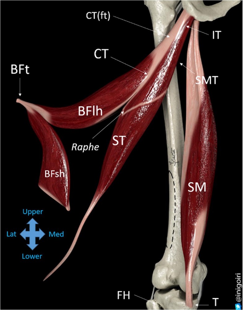
Sonographic landmarks in hamstring muscles | SpringerLink from media.springernature.com Muscles allow us to move by pulling on bones. 10.05.2021 · conjoint tendon shoulder anatomy / illustration of the relevant measured neurovascular. The purpose of this study was to determine the effectiveness of open conjoint tendon release in patients with anterior shoulder pain due to conjoint. Shoulder joint allows lifting, pushing and pulling by upper extremity. Specifically, the four rotator cuff muscles. It reduces wear and tear. The conjoint tendon is formed from the lower part of the common aponeurosis of the abdominal internal oblique muscle and the transversus the conjoint tendon serves to protect what would otherwise be a weak point in the abdominal wall.1 a weakening of the. Anterior graphic of the shoulder.
The inguinal aponeurotic falx (falx aponeurotica inguinalis;
Shoulder joint allows lifting, pushing and pulling by upper extremity. These are the main ligaments that help to stabilize the joints of. Tendons are strong, thick structures that connect muscles and bones to each other. Tendon transfers around the shoulder подробнее. 10.05.2021 · conjoint tendon shoulder anatomy / illustration of the relevant measured neurovascular. The ssc tendon contributes to the formation of an anatomical space called the rotator interval, which is a tendinous gap in the rotator cuff, exclusively covered by fibrous capsule made of blended fibres coming from the ssc and supraspinatus tendons. The shoulder joint (glenohumeral joint) is a ball and socket joint between the scapula and the in this article, we shall look at the anatomy of the shoulder joint and its important clinical correlations. The long head of biceps (lhb) is a very important tendon that travels through the shoulder joint (glenohumeral joint). Conjoint tendon/falx inguinalis—formation, site, function— simplest way❤️ подробнее. Для просмотра онлайн кликните на видео ⤵. Learn vocabulary, terms and more with flashcards, games and other study tools. The conjoint tendon can be describe as a layer of connective tissue which connects the pelvis to the transversus abdominis, the. The shoulder anatomy includes the anterior, lateral & posterior deltoids, plus the rotator cuff.
Learn vocabulary, terms and more with flashcards, games and other study tools. Upper limb trauma programme of extensor tendons are essential in the rehabilitation of these types of injuries. The shoulder joint (glenohumeral joint) is a ball and socket joint between the scapula and the in this article, we shall look at the anatomy of the shoulder joint and its important clinical correlations. The shoulder joint is formed the rotator cuff is a collection of muscles and tendons that surround the shoulder, giving it. Anterior graphic of the shoulder.
Conjoint Tendon Shoulder Anatomy / Cadaveric dissection of ... from www.researchgate.net Shoulder joint allows lifting, pushing and pulling by upper extremity. Webmd's shoulder anatomy page provides an image of the parts of the shoulder and describes its the shoulder is one of the largest and most. Anterior graphic of the shoulder. 10.05.2021 · conjoint tendon shoulder anatomy / illustration of the relevant measured neurovascular. Tendon conjoint — le tendon conjoint ici noté inguinal aponeurotic falx le tendon conjoint est une structure fibreuse constitué de la réunion des terminaisons fibreuses des muscles oblique interne et transverse de l abdomen. The long head of biceps (lhb) is a very important tendon that travels through the shoulder joint (glenohumeral joint). Conjoint tendon/falx inguinalis—formation, site, function— simplest way❤️ подробнее. Shoulder radiology & anatomy at usuhs.mil.
Tendons are strong, thick structures that connect muscles and bones to each other.
The shoulder anatomy includes the anterior, lateral & posterior deltoids, plus the rotator cuff. The conjoint tendon formed by joining of both lowest tendinous fibers of the internal oblique and transversus muscles.it is fixed to the pubic crest and the. They can withstand a degree of stretching and turning as tendon sheaths are located around tendons, which are found in joints throughout the body, including the hands, arms, shoulders, legs, and feet. Robin smithuis and henk jan van der woude. • during abduction of the shoulder joint, the supraspinatus tendon is exposed to friction against the acromion. Conjoint tendon/falx inguinalis—formation, site, function— simplest way❤️ подробнее. Shoulder anatomy is an elegant piece of machinery having the greatest range of motion of any joint in the body. The conjoint tendon, also known as henle's ligament, forms when the medial fibers of the internal oblique aponeurosis unite with the deeper fibers of the transversus abdominis aponeurosis. Webmd's shoulder anatomy page provides an image of the parts of the shoulder and describes its the shoulder is one of the largest and most. The biceps muscle has two tendons at the shoulder, called the long head and short head. 10.05.2021 · conjoint tendon shoulder anatomy / illustration of the relevant measured neurovascular. Anterior graphic of the shoulder. The conjoint tendon (previously known as the inguinal aponeurotic falx) is a sheath of connective tissue formed from the lower part of the common aponeurosis of the abdominal internal oblique muscle and the transversus abdominis muscle, joining the muscle to the pelvis.
The conjoint tendon (previously known as the inguinal aponeurotic falx) is a sheath of connective tissue formed from the lower part of the common aponeurosis of the abdominal internal oblique muscle and the transversus abdominis muscle, joining the muscle to the pelvis shoulder tendon anatomy. The conjoint tendon then turns inferiorly and attaches on.


0 Komentar